The link between COVID-19 and heel pain

Since the COVID-19 pandemic began in 2020, the number of patients reporting heel pain has risen. The condition is now so common it has come to be known as “pandemic foot”. Is there really a link between COVID-19 and heel pain, and if so, what treatment options are available?

What is plantar fasciitis?

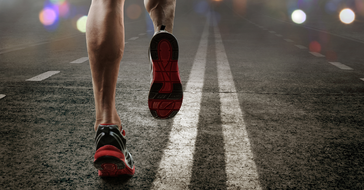
While “pandemic foot” might be a catchy name, the correct medical term is plantar heel pain, or plantar fasciitis. This condition presents as pain felt on the bottom of the foot around the heel and arch. It is an overuse condition often associated with runners, especially those over the age of 40.

Excessive pressure on the foot, along with a tight calf or Achilles tendon, can cause inflammation of the plantar fascia, the thick band of tissue on the bottom of the foot connecting the heel to the toes.

The pain is commonly felt during the first step, as well as during weight-bearing tasks, particularly after periods of rest.1 Patients often report the pain at its worst as they take their first steps of the day after getting out of bed. It typically decreases as the calf and Achilles tendon become looser during activity, only to return the following day after things have tightened up again during the night.

Does COVID-19 cause plantar fasciitis?

There is no current evidence to suggest there is a direct link between COVID-19 and heel pain. Instead, the rise in plantar fasciitis is more likely to be due to the changes in our daily lives the pandemic has brought about.

Gym attendances have declined since the beginning of the pandemic, with outdoor running and walking becoming more popular instead. More running and walking mean more stress on the plantar fascia, which, due to an increase in flexible working, can be exacerbated by more time spent walking around at home in bare feet, slippers, or flip-flops.

Without the additional support that a heeled shoe can provide, like those typically worn in office environments, the foot spends more time in a flat position, which, for extended periods, can put additional strain on the fascia. Add to this stiff muscles and tendons from running, and you have a recipe for plantar fasciitis. This is the indirect link between COVID-19 and heel pain.

How can heel pain be treated?

There are a number of conservative treatment options for plantar fasciitis. They range from relatively simple orthotics to more advanced rehabilitation devices.

Taping for heel pain

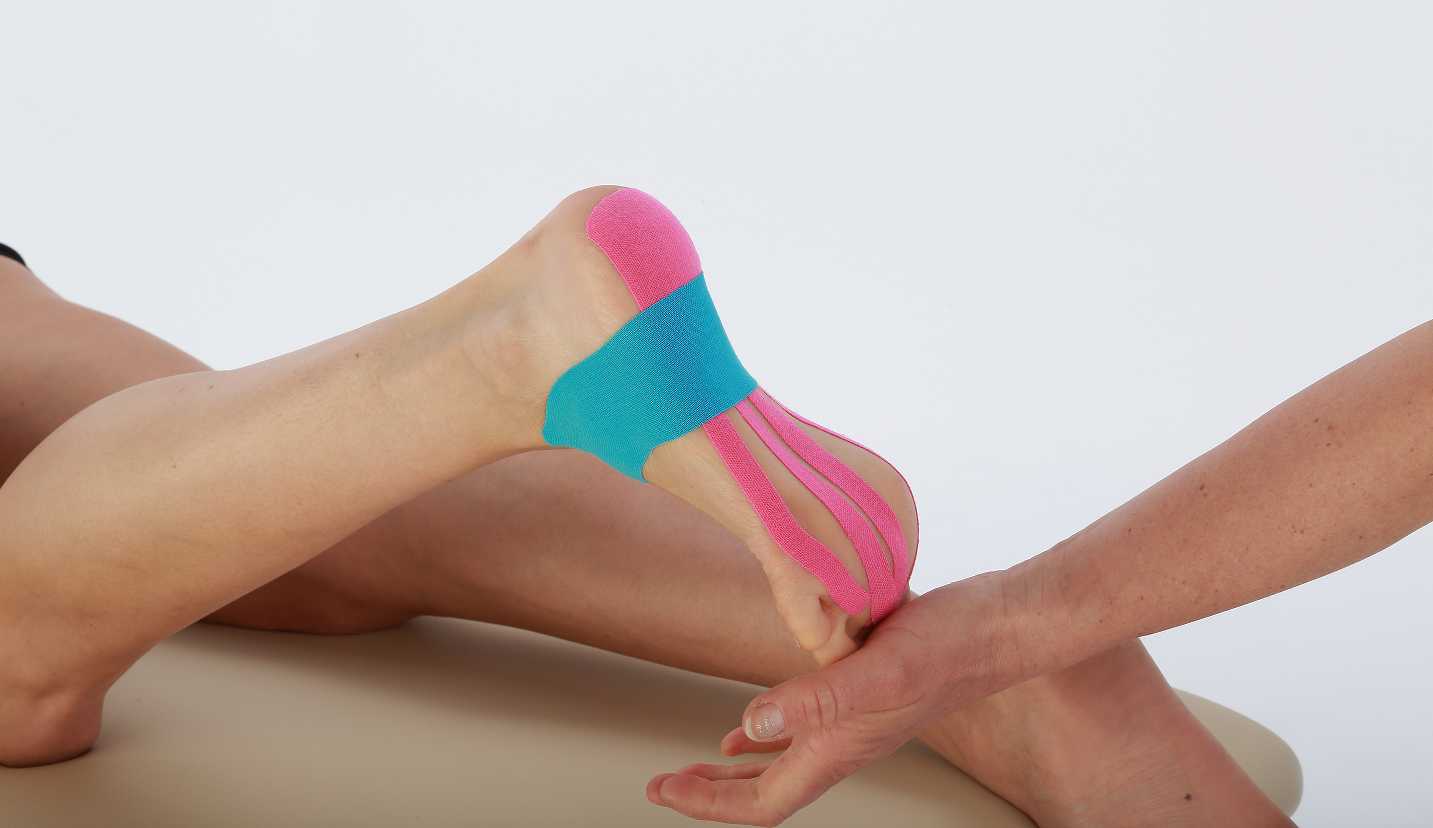
Physio tape (also known as kinesiology tape) like Chatt-Tape is elastic adhesive tape that can be applied to parts of the body to aid healing and recuperation of the soft tissue.2

Tape can be applied to the heel, ankle, and underside of the foot to release tension in the plantar fascia as well as stabilize it. A study by Tezel et al. (2020) showed that kinesiology tape provided pain relief and improved quality of life for patients with plantar fasciitis, as well as improved functionality.3

Chatt-Tape plantar fasciitis
Aircast AirHeel and Dorsal Night Splint

Bracing for heel pain

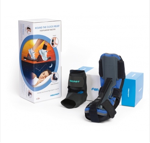
Plantar fasciitis can be relieved by wearing an orthotic during the night to help reduce the tightening of the calf muscles and Achilles tendon.4 One such device is Aircast’s Dorsal Night Splint; this product is worn while the patient sleeps, to maintain the position of the foot at 90°, thereby helping to stretch the calf and Achilles tendon.

Another type of foot orthosis for plantar fasciitis is a pneumatic ankle brace. Also from Aircast, the AirHeel is designed to treat plantar fasciitis, Achilles tendonitis, and heel pain. Using two interconnected aircells located under the foot arch and in the back of the heel, the brace applies pulsating compression with every step to help reduce swelling and discomfort and enhance circulation.

Kavros’s 2005 study showed that patients with higher plantar fasciitis pain experience faster relief with the Airheel than with a shoe insert.5

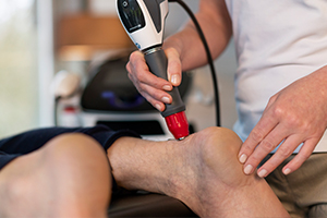
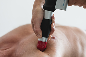
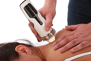
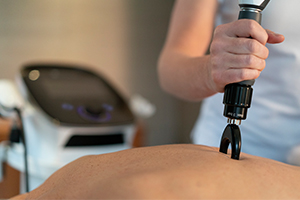
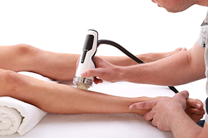
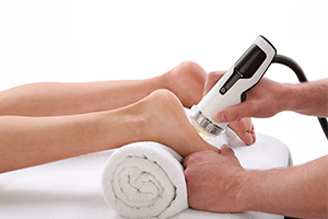
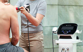
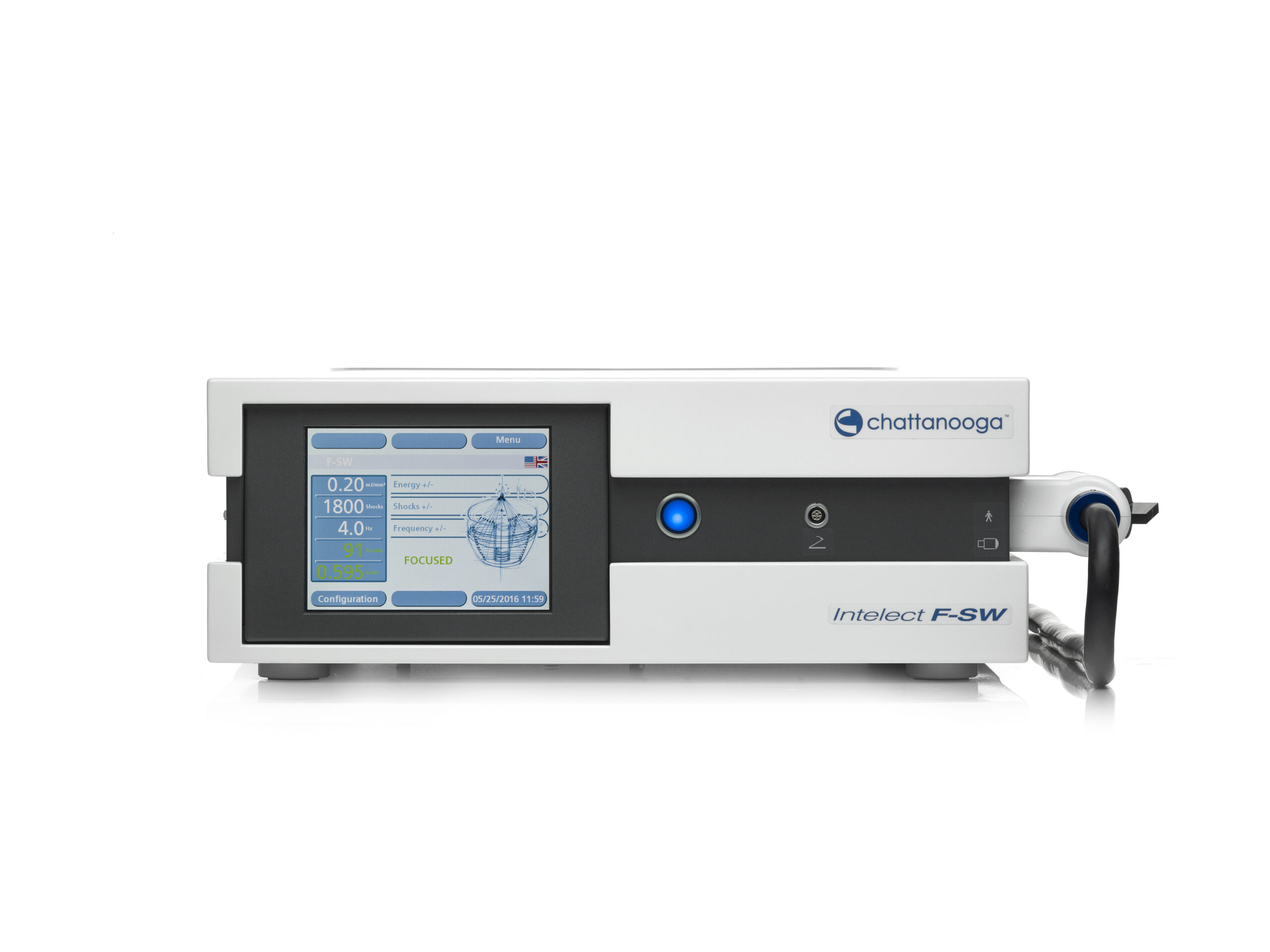
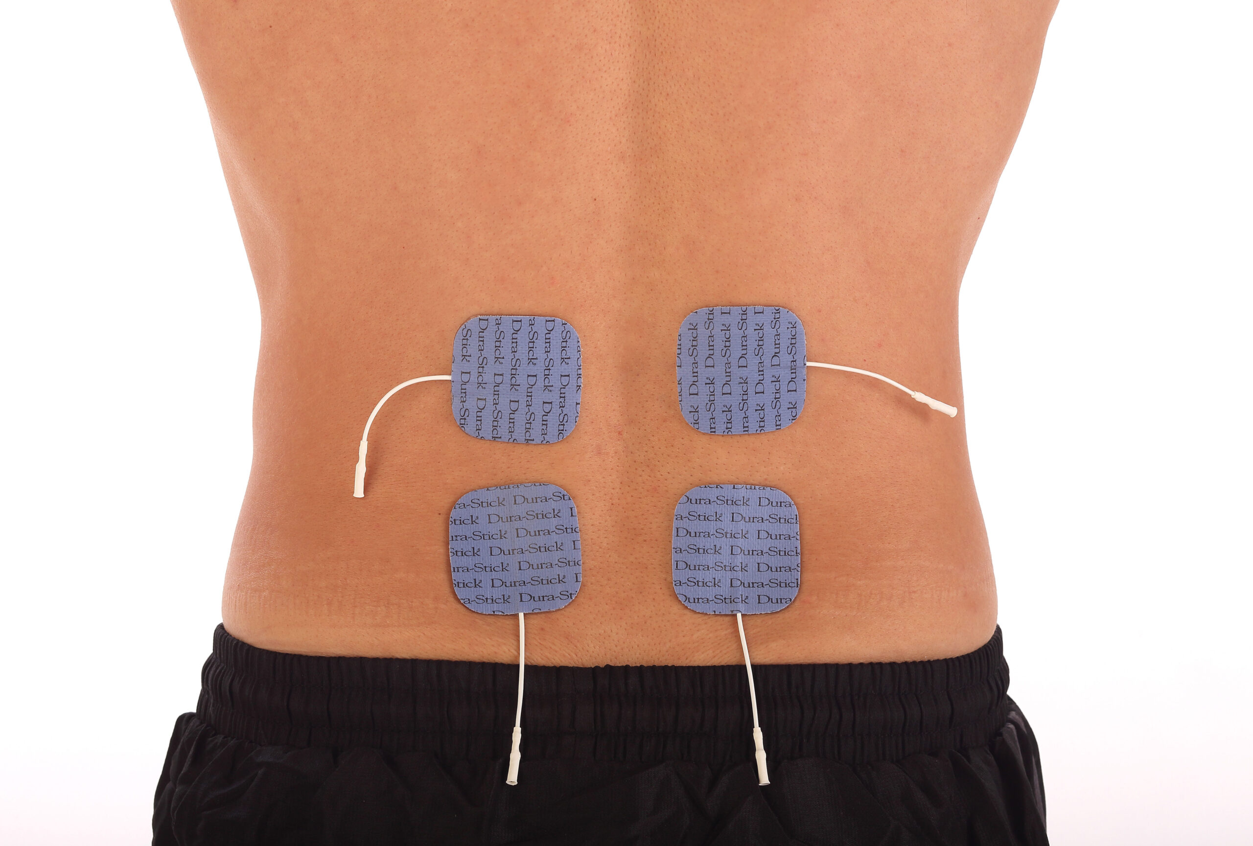
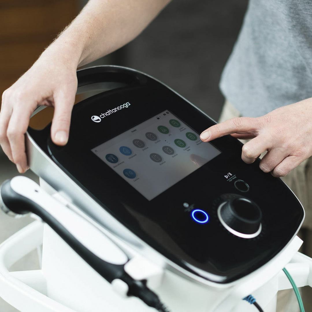
Shock wave therapy for heel pain

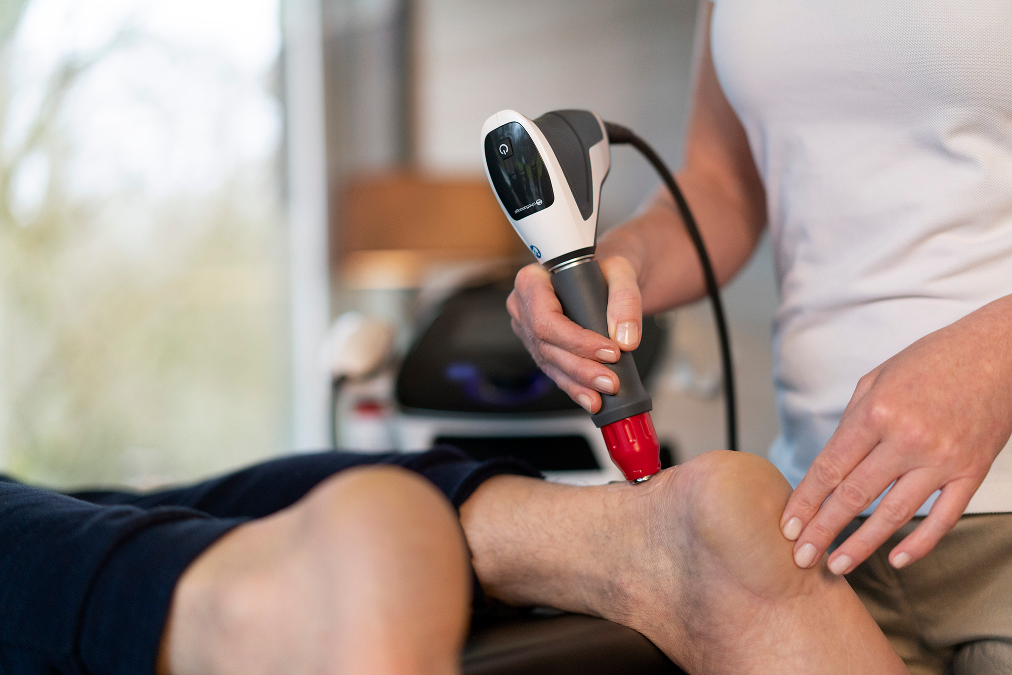
Shock wave therapy is an electronic modality that uses acoustic waves to stimulate the body on a cellular level for healing purposes. Generally divided into focused shock wave (F-SW) and radial pressure wave (RPW) therapy, shock wave therapy has been shown to be a clinically proven treatment option for plantar fasciitis, especially when treatments like taping have not been successful.1

In a 2022 study by Wheeler et al., RPW treatment provided significant improvement of pain and function in patients with chronic plantar fasciopathy.6

Intelect 2 RPW
LightForce laser therapy

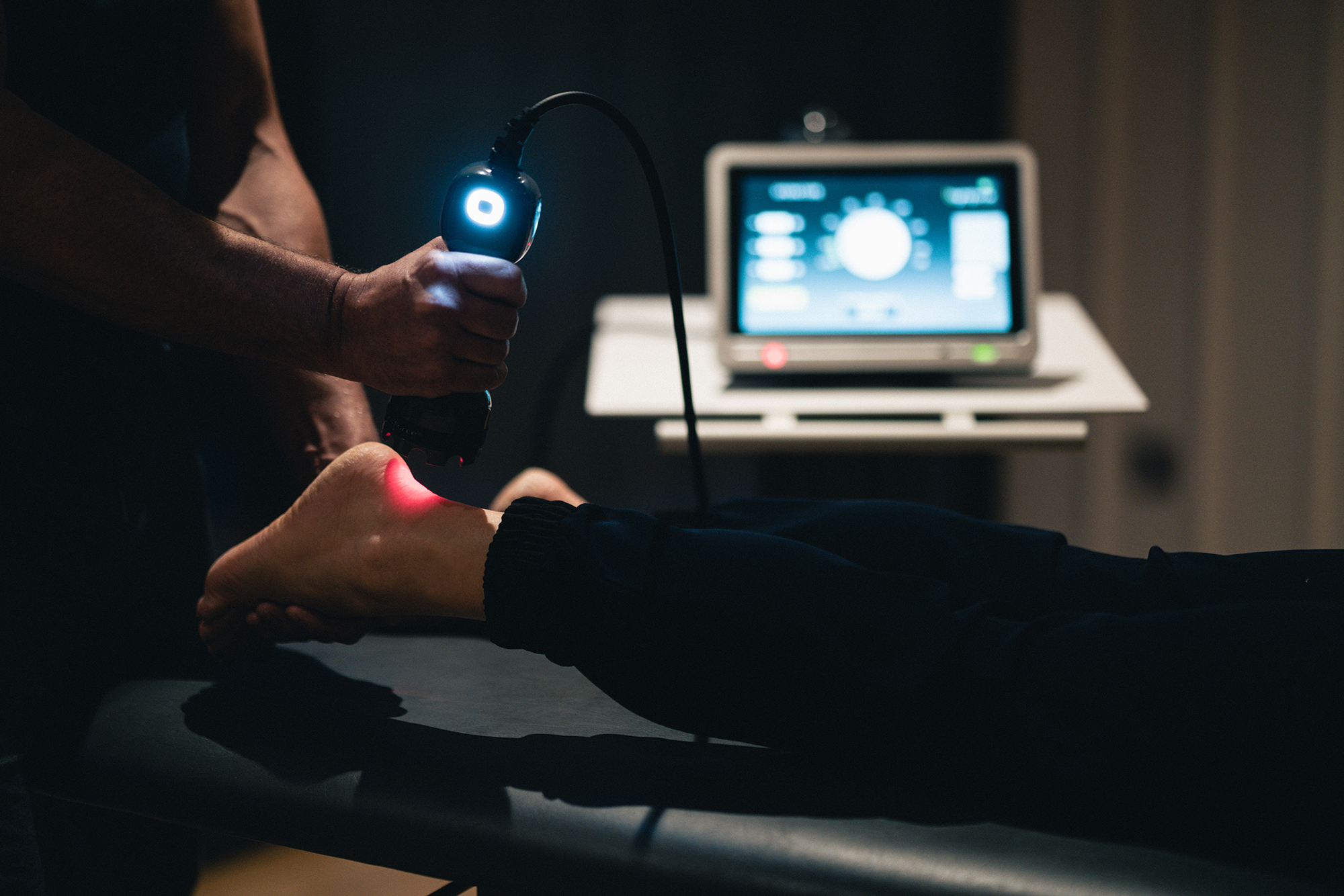
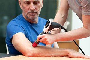
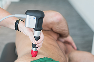
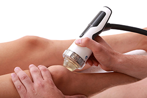
High power laser therapy for heel pain

High power laser therapy, like that offered by LightForce, uses the energy of focused light to trigger the body’s natural healing processes, thereby speeding recovery.

Ordahan et al.’s 2018 study demonstrated that high power laser therapy provided improvement of pain and function in patients with plantar fasciitis.7

Combining laser therapy with shock wave therapy has shown to be even more effective.8

To learn more about products for heel pain, visit enovis-medtech.eu

References

  1. Morrissey, D., Cotchett, M., Said J’Bari, A., Prior, T., Griffiths, I. B., Rathleff, M. S., Gulle, H., Vicenzino, B., & Barton, C. J. (2021). Management of plantar heel pain: a best practice guide informed by a systematic review, expert clinical reasoning and patient values. British journal of sports medicine, 55(19), 1106–1118.
  2. Homayouni, K., et al. (2013). Comparison between kinesio taping and physiotherapy in the treatment of de Quervain’s disease. J. Musculoskelet. Res. 16(4).
  3. Tezel, N., Umay, E., Bulut, M., Cakci, A (2020). Short-Term Efficacy of Kinesiotaping versus Extracorporeal Shockwave Therapy for Plantar Fasciitis: A Randomized Study. Saudi J Med Med Sci. Sep-Dec;8(3):181-187.
  4. Powell, M., Post, W. R., Keener, J., & Wearden, S. (1998). Effective treatment of chronic plantar fasciitis with dorsiflexion night splints: a crossover prospective randomized outcome study. Foot & ankle international, 19(1), 10–18.
  5. Kavros, S. J. (2005). The efficacy of a pneumatic compression device in the treatment of plantar fasciitis. Journal of applied biomechanics, 21(4), 404–413.
  6. Wheeler, P. C., Dudson, C., & Calver, R. (2022). Radial Extracorporeal Shockwave Therapy (rESWT) is not superior to “minimal-dose” rESWT for patients with chronic plantar fasciopathy; a double-blinded randomised controlled trial. Foot and ankle surgery : official journal of the European Society of Foot and Ankle Surgeons, 28(8), 1356–1365.
  7. Ordahan, B., Karahan, A. Y., & Kaydok, E. (2018). The effect of high-intensity versus low-level laser therapy in the management of plantar fasciitis: a randomized clinical trial. Lasers in medical science, 33(6), 1363–1369.
  8. Takla, M. K. N., & Rezk, S. S. R. (2019). Clinical effectiveness of multi-wavelength photobiomodulation therapy as an adjunct to extracorporeal shock wave therapy in the management of plantar fasciitis: a randomized controlled trial. Lasers in medical science, 34(3), 583–593.

What can shock wave therapy be used to treat?

Focused shock wave (F-SW) therapy and radial pressure wave (RPW) therapy have been shown to stimulate the body’s natural healing process, with positive effects on bone and tendon repair, as well as tissue regeneration26. But how can these effects be applied practically to treat patients?

COMMON INDICATIONS FOR SHOCK WAVE THERAPY

Tendon pathologies – hamstrings, Achilles tendon, patellar tendon, shoulder ‘rotator cuff’

Tendon pathologies – hamstrings, Achilles tendon, patellar tendon, shoulder ‘rotator cuff’

F-SW and RPW While it was a long-held belief that chronic tendons were not capable of repair, studies have shown that F-SW is effective in significantly stimulating the growth indicators associated with tendon, bone, and tendon-bone interface1,2. Positive outcomes have also been recorded with RPW treatment3.  

Frozen shoulder

F-SW and RPW Frozen shoulder is an idiopathic and progressive disease, identified by pain and decreased range of motion (ROM) of the shoulder and shoulder joint capsule fibrosis. The use of F-SW seems to have positive effects on treatment, quicker return to daily activities, and quality-of-life improvement on frozen shoulder4. RPW has also been shown to be effective5.

Calcifications F-SW and RPW Both F-SW and RPW can provide positive results in reducing calcification, with improvements recorded in shoulder pain, range of motion, and function, while combining the two has been shown to provide even better results6.  

Tennis elbow

F-SW and RPW  Tennis elbow, or lateral epicondylitis, is a common source of pain among manual workers. It has been demonstrated that F-SW was more effective than ultrasound therapy for improving pain and grip strength when treating tennis elbow, and also yielded better subjective evaluation7. Additionally, RPW therapy has been shown to yield higher improvements than steroid injections in treating lateral epicondylitis8.

Carpal Tunnel Syndrome F-SW and RPW Positive outcomes have been observed for pain symptoms, functional outcomes, and median nerve activity9.  

Chronic neck pain F-SW and RPW F-SW is more effective than ultrasound for improvement of myofascial pain syndrome, and equally effective as dry needling and laser therapy. It is also less invasive and less prone to adverse effects or allergic reactions than those conventional therapies10. Trigger point treatment with RPW used in combination with physical therapy has also been shown to relieve neck and shoulder pains11. However, it is important to mention that the anterior cervical area is contraindicated for localized shock wave therapy, and that only experienced clinicians should consider such treatments, as care must be taken to avoid neurovascular bundles.

Low back pain

F-SW and RPW Studies have shown that RPW added to conventional physiotherapy and core stability exercises has a significant effect on the reduction of chronic low back pain and the improvement of functional condition compared to a conventional physiotherapy program12. F-SW has also proven effective13.

Muscle hypertonia F-SW and RPW It has been demonstrated that both F-SW and RPW are effective in reducing muscle spasticity and improving motor recovery after stroke14, while RPW has also shown positive results in reducing pain and muscle tone in multiple sclerosis patients as part of a rehabilitation program15.  

Hip pain

F-SW and RPW While low-energy shock wave interventions cannot be used to treat hip conditions such as avascular necrosis or intracapsular pathology, RPW has been shown to be effective in treating greater trochanter hip pain16,17. F-SW appears to be effective for aiding in pain relief and functional recovery in patients with osteonecrosis of the hip18.

Knee osteoarthritis

F-SW F-SW has been proven effective for improving pain and function in knee OA, with medium energy values having significantly greater effect than low energy19,20.  

Shin pain (‘splints’) F-SW and RPW Traditional treatment of medial tibial stress syndrome (MTSS) is generally lengthy, associated with frequent recurrences, and in some cases, an unacceptable degree of improvement. In one study, a single application of F-SW treatment in combination with a specific exercise programme accelerated clinical and functional recovery from MTSS21. Adding RPW to standard home exercise therapy has been shown to offer significantly more improvement of pain, degree of recovery, and return to sports22.

Plantar fasciitis

F-SW and RPW F-SW has been shown effective in reducing heel pain associated with chronic plantar fasciitis23. RPW therapy has been demonstrated to improve pain, function, and quality of life in patients with recalcitrant plantar fasciitis24.  

Erectile dysfunction F-SW The number of studies of low-intensity focused shock wave therapy for erectile dysfunction (ED) have increased dramatically in recent years, with results indicating F-SW significantly improves ED and the efficacy can last up to 3 months and more. Furthermore, it may have the potential to be the first-choice non-invasive treatment for patients with ED25.  

Wound healing F-SW It has been well established that, through the principle of ‘mechanotransduction’ (the process by which a mechanical stimulus is converted into a set of biochemical reactions and a cellular response), F-SW can positively influence the chain of biological reactions that lead to tissue regeneration and healing26.  

Diabetic foot F-SW As with wound healing in general, F-SW as an adjunct to standard wound care has been shown to significantly reduce the size and the healing time for wounds associated with chronic diabetic foot ulcers27.  

Cellulite RPW RPW therapy is well established in the aesthetics market, having been shown to be an effective treatment for improving the appearance of cellulite, with significant improvement recorded in skin firmness and structure28.  

REFERENCES

  1. Notarnicola A, Moretti B. The biological effects of extracorporeal shock wave therapy (eswt) on tendon tissue. Muscles Ligaments Tendons J. 2012 Jun 17;2(1):33-7.
  2. Waugh CM, Morrissey D, Jones E, Riley GP, Langberg H, Screen HR. In vivo biological response to extracorporeal shockwave therapy in human tendinopathy. Eur Cell Mater. 2015 May 15;29:268-80; discussion 280.
  3. Rompe JD, Maffulli N. Repetitive shock wave therapy for lateral elbow tendinopathy (tennis elbow): a systematic and qualitative analysis. Br Med Bull. 2007;83:355-78.
  4. Vahdatpour B, Taheri P, Zade AZ, Moradian S. Efficacy of extracorporeal shockwave therapy in frozen shoulder. Int J Prev Med. 2014 Jul;5(7):875-81.
  5. Hussein AZ, Donatelli RA. () The efficacy of radial extracorporeal shockwave therapy in shoulder adhesive capsulitis: a prospective, randomised, double-blind, placebo-controlled, clinical study. European Journal of Physiotherapy. 2016; 18:1, 63-76,
  6. Abo Al-Khair MA, El Khouly RM, Khodair SA, Al Sattar Elsergany MA, Hussein MI, Eldin Mowafy ME. Focused, radial and combined shock wave therapy in treatment of calcific shoulder tendinopathy. Phys Sportsmed. 2020 Dec 6:1-8.
  7. Yan C, Xiong Y, Chen L, Endo Y, Hu L, Liu M, Liu J, Xue H, Abududilibaier A, Mi B, Liu G. A comparative study of the efficacy of ultrasonics and extracorporeal shock wave in the treatment of tennis elbow: a meta-analysis of randomized controlled trials. J Orthop Surg Res. 2019 Aug 6;14(1):248.
  8. Beyazal MS et al. (Turkey). Comparison of the effectiveness of local corticosteroid injection and extracorporeal shock wave therapy in patients with lateral epicondylitis. J Phys Ther Sci. 2015 Dec;27(12):3755-8.
  9. Kim JC, Jung SH, Lee SU, Lee SY. Effect of extracorporeal shockwave therapy on carpal tunnel syndrome: A systematic review and meta-analysis of randomized controlled trials. Medicine (Baltimore). 2019 Aug;98(33):e16870.
  10. Zhang Q, Fu C, Huang L, Xiong F, Peng L, Liang Z, Chen L, He C, Wei Q. Efficacy of Extracorporeal Shockwave Therapy on Pain and Function in Myofascial Pain Syndrome of the Trapezius: A Systematic Review and Meta-Analysis. Arch Phys Med Rehabil. 2020 Aug;101(8):1437-1446.
  11. Damian M, Zalpour C. Trigger point treatment with radial shock waves in musicians with nonspecific shoulder-neck pain: data from a special physio outpatient clinic for musicians. Med Probl Perform Art. 2011 Dec;26(4):211-7.
  12. Walewicz K, Taradaj J, Dobrzyński M, Sopel M, Kowal M, Ptaszkowski K, Dymarek R. Effect of Radial Extracorporeal Shock Wave Therapy on Pain Intensity, Functional Efficiency, and Postural Control Parameters in Patients with Chronic Low Back Pain: A Randomized Clinical Trial. J Clin Med. 2020 Feb 19;9(2):568.
  13. Hong JO, Park JS, Jeon DG, Yoon WH, Park JH. Extracorporeal Shock Wave Therapy Versus Trigger Point Injection in the Treatment of Myofascial Pain Syndrome in the Quadratus Lumborum. Ann Rehabil Med. 2017 Aug;41(4):582-588.
  14. Dymarek R, Ptaszkowski K, Ptaszkowska L, Kowal M, Sopel M, Taradaj J, Rosińczuk J. Shock Waves as a Treatment Modality for Spasticity Reduction and Recovery Improvement in Post-Stroke Adults – Current Evidence and Qualitative Systematic Review. Clin Interv Aging. 2020 Jan 6;15:9-28.
  15. Marinelli L, Mori L, Solaro C, Uccelli A, Pelosin E, Currà A, Molfetta L, Abbruzzese G, Trompetto C. Effect of radial shock wave therapy on pain and muscle hypertonia: a double-blind study in patients with multiple sclerosis. Mult Scler. 2015 Apr;21(5):622-9.
  16. Rompe JD, Segal NA, Cacchio A, Furia JP, Morral A, Maffulli N. Home training, local corticosteroid injection, or radial shock wave therapy for greater trochanter pain syndrome. Am J Sports Med. 2009 Oct;37(10):1981-90.
  17. Furia JP, Rompe JD, Maffulli N. Low-energy extracorporeal shock wave therapy as a treatment for greater trochanteric pain syndrome. Am J Sports Med. 2009 Sep;37(9):1806-13.
  18. Zhao W, Gao Y, Zhang S, Liu Z, He L, Zhang D, Li W, Meng Q. Extracorporeal shock wave therapy for bone marrow edema syndrome in patients with osteonecrosis of the femoral head: a retrospective cohort study. J Orthop Surg Res. 2021 Jan 7;16(1):21.
  19. Lee JK, Lee BY, Shin WY, An MJ, Jung KI, Yoon SR. Effect of Extracorporeal Shockwave Therapy Versus Intra-articular Injections of Hyaluronic Acid for the Treatment of Knee Osteoarthritis. Ann Rehabil Med. 2017 Oct;41(5):828-835.
  20. Kim JH, Kim JY, Choi CM, Lee JK, Kee HS, Jung KI, Yoon SR. The Dose-Related Effects of Extracorporeal Shock Wave Therapy for Knee Osteoarthritis. Ann Rehabil Med. 2015 Aug;39(4):616-23.
  21. Gomez Garcia S, Ramon Rona S, Gomez Tinoco MC, Benet Rodriguez M, Chaustre Ruiz DM, Cardenas Letrado FP, Lopez-Illescas Ruiz Á, Alarcon Garcia JM. Shockwave treatment for medial tibial stress syndrome in military cadets: A single-blind randomized controlled trial. Int J Surg. 2017 Oct;46:102-109.
  22. Rompe JD, Cacchio A, Furia JP, Maffulli N. Low-energy extracorporeal shock wave therapy as a treatment for medial tibial stress syndrome. Am J Sports Med. 2010 Jan;38(1):125-32.
  23. Gollwitzer H et al. (Technische Universität München, Munich, Germany). Clinically relevant effectiveness of focused extracorporeal shock wave therapy in the treatment of chronic plantar fasciitis: a randomized, con­trolled multicenter study. J Bone Joint Surg Am. 2015 May 6;97(9):701-8.
  24. Gerdesmeyer L, Frey C, Vester J, Maier M, Weil L Jr, Weil L Sr, Russlies M, Stienstra J, Scurran B, Fedder K, Diehl P, Lohrer H, Henne M, Gollwitzer H. Radial extracorporeal shock wave therapy is safe and effective in the treatment of chronic recalcitrant plantar fasciitis: results of a confirmatory randomized placebo-controlled multicenter study. Am J Sports Med. 2008 Nov;36(11):2100-9.
  25. Lu Z, Lin G, Reed-Maldonado A, Wang C, Lee YC, Lue TF. Low-intensity Extracorporeal Shock Wave Treatment Improves Erectile Function: A Systematic Review and Meta-analysis. Eur Urol. 2017 Feb;71(2):223-233.
  26. d’Agostino MC, Craig K, Tibalt E, Respizzi S. Shock wave as biological therapeutic tool: From mechanical stimulation to recovery and healing, through mechanotransduction. Int J Surg. 2015 Dec;24(Pt B):147-53.
  27. Omar MT, Alghadir A, Al-Wahhabi KK, Al-Askar AB. Efficacy of shock wave therapy on chronic diabetic foot ulcer: a single-blinded randomized controlled clinical trial. Diabetes Res Clin Pract. 2014 Dec;106(3):548-54.
  28. Russe-Wilflingseder K, Russe E, Vester JC, Haller G, Novak P, Krotz A. Placebo controlled, prospectively randomized, double-blinded study for the investigation of the effectiveness and safety of the acoustic wave therapy (AWT(®)) for cellulite treatment. J Cosmet Laser Ther. 2013 Jun;15(3):155-62.

What are the effects of shock wave therapy?

Shock wave therapy – a term that continues to be used to encompass both focused shock wave and radial pressure wave therapies – can be used to stimulate the body’s natural healing process, with positive effects demonstrated on bone and tendon repair and tissue regeneration1. But how does this work?

Both focused shock waves and radial pressure waves influence cellular activity by mechanotransduction, where the mechanical energy of an acoustic wave is converted into biochemical energy in the cell and extracellular matrix1.

With focused shock wave (F-SW) therapy, this is primarily achieved through the effects of cavitation, whereas radial pressure wave (RPW) therapy uses pressure waves to cause cellular change.

What is cavitation?

Cavitation occurs during the tensile phase of a shock wave. The negative pressure created by the wave generates gas-filled bubbles in water at room temperature, which are normally only seen in boiling water.

When these bubbles are concentrated in a small area, such as in F-SW therapy, they generate secondary pressure waves in a process known as “stable cavitation.” When the bubbles burst, they release energy at the focal point of the application. This “microtrauma” stimulates the body’s self-healing ability and leads to tissue regeneration2,3.

If the bubbles are encouraged to expand, they will continue to store energy until they become so large that they implode, releasing a large amount of energy in a microjet. This is known as “unstable cavitation,” and can be used to break down kidney stones4,5.

While some cavitation also occurs during RPW therapy, the level is only superficial, and not thought to be significant.

How shock wave therapy helps the body to heal

F-SW and RPW therapies are pro-inflammatory modalities which can be used to “reboot” the healing process in stalled, chronic conditions1.

The normal healing process of the body occurs in four stages. First the body responds to injury by bleeding, which is then followed by inflammation. Within a few hours to days, the third stage begins, known as proliferation, where new tissue is created to rebuild the wound. Finally, after a few weeks, the remodelling stage starts, during which the wound fully closes.

Of all these stages of the healing cascade, the most important is inflammation. Without it, proliferation and remodelling will not take place. In chronic conditions, something has gone wrong during the later proliferation and remodelling stages of the normal healing process, preventing it from progressing. Like a computer with a serious error, this process needs to be rebooted for progress to be made by using the pro-inflammatory action of F-SW or RPW therapy.

Other physiological effects of shock wave therapy

Along with its influence on inflammation, shock wave therapy has been shown to have a number of other physical effects on the body:

New blood vessel formation

Due to microtraumas caused by shock wave therapy, there is a significant increase in the expression of growth factors such as eNOS, VEGF, PCNS and BMP. These growth factors are involved in the process of neovascularization, where arterioles are stimulated to form and grow, which has a positive effect on improved blood supply, bone and tendon repair, and tissue regeneration6.

Collagen production

Shock wave therapy stimulates procollagen synthesis, necessary for the repair of damaged musculoskeletal and ligament structures. It forces the newly created collagen fibers into a longitudinal structure, making the newly formed tendon fibers denser and stiffer, with a firmer structure7.  

Tenocyte proliferation

Shock wave therapy leads to an increase in transforming growth factor beta 1 (TGF-β 1), which is known to regulate tendon repair8.

Fibrotic tissue and scar remodelling

Shock wave therapy alters the expression of fibrosis-related molecules in fibroblasts, which affects scar remodelling and resorption1.

Osteoblast and osteoclast activity

Alongside TGF-β 1, fibroblast growth factor 2 (FGF2) is also increased by shock wave therapy, and together they are influential in bone healing1,9.

Analgesia

RPW therapy has a confirmed effect in improving the symptoms of pain10.

Reducing hypertonia in spastic muscles

Both F-SW and RPW have been proven effective here11, with RPW particularly effective in aiding increased range of movement12.

To learn more about the effects and applications of shock wave therapy:

References

  1. d’Agostino MC, Craig K, Tibalt E, Respizzi S. Shock wave as biological therapeutic tool: From mechanical stimulation to recovery and healing, through mechanotransduction. Int J Surg. 2015 Dec;24(Pt B):147-53. 
  2. Ogden JA, Tóth-Kischkat A, Schultheiss R. Principles of shock wave therapy. Clin Orthop Relat Res. 2001 Jun;(387):8-17.
  3. Császár NB, Angstman NB, Milz S, Sprecher CM, Kobel P, Farhat M, Furia JP, Schmitz C. Radial Shock Wave Devices Generate Cavitation. PLoS One. 2015 Oct 28;10(10):e0140541.
  4. Chaussy C, Brendel W, Schmidt E. Extracorporeally induced destruction of kidney stones by shock waves. Lancet 2:1265–1268, 1980.
  5. Streem SB. Contemporary clinical practice of shock wave lithotripsy: a reevaluation of contraindications. J Urol. 1997 Apr;157(4):1197-203.
  6. Wang CJ, Wang FS, Yang KD. Biological mechanism of musculoskeletal shockwaves. ISMST Newsletter 2006, 1 (I), 5-11.
  7. Vetrano M, d’Alessandro F, Torrisi MR, Ferretti A, Vulpiani MC, Visco V. Extracorporeal shock wave therapy promotes cell proliferation and collagen synthesis of primary cultured human tenocytes. Knee Surg Sports Traumatol Arthrosc. 2011 Dec;19(12):2159-68.
  8. Berta L, Fazzari A, Ficco AM, Enrica PM, Catalano MG, Frairia R. Extracorporeal shock waves enhance normal fibroblast proliferation in vitro and activate mRNA expression for TGF-beta1 and for collagen types I and III. Acta Orthop. 2009 Oct;80(5):612-7.
  9. Frairia R, Berta L. Biological effects of extracorporeal shock waves on fibroblasts. A review. Muscles Ligaments Tendons J. 2012 Apr 1;1(4):138-47.
  10. Rompe JD, Hope C, Küllmer K, Heine J, Bürger R. Analgesic effect of extracorporeal shock-wave therapy on chronic tennis elbow. J Bone Joint Surg Br. 1996 Mar;78(2):233-7.
  11. Dymarek R, Ptaszkowski K, Ptaszkowska L, Kowal M, Sopel M, Taradaj J, Rosińczuk J. Shock Waves as a Treatment Modality for Spasticity Reduction and Recovery Improvement in Post-Stroke Adults – Current Evidence and Qualitative Systematic Review. Clin Interv Aging. 2020 Jan 6;15:9-28.
  12. Marinelli L, Mori L, Solaro C, Uccelli A, Pelosin E, Currà A, Molfetta L, Abbruzzese G, Trompetto C. Effect of radial shock wave therapy on pain and muscle hypertonia: a double-blind study in patients with multiple sclerosis. Mult Scler. 2015 Apr;21(5):622-9.

What is the difference between focused shock wave (FSW) and radial pressure wave (RPW) therapy?

Shock wave therapy is not a new modality; in its modern electronic form it has been around since the early 1980s, where it was first shown to have beneficial effects on treating kidney and biliary stones, and later on bone and wound healing.

However, despite now being an established medical intervention, questions still persist around the specifics of shock wave therapy. This article explains the difference between focused shock wave (F-SW) and radial pressure wave (RPW) therapy.

What are shock waves?

A shock wave is defined as a moving sound source travelling at more than the speed of sound. A stationary sound wave emits acoustic pressure waves that are evenly distributed in all directions. However, when the sound source is moving, the sound waves in front are compressed, and if the source moves faster than the speed of sound, the compressed waves overlap and create a shock wave, which is heard as a sonic boom.

It was noted that shock waves travel much better through water than they do through air, and that they could affect the body on a cellular level, thereby stimulating its intrinsic healing mechanism. This led to efforts to harness this phenomenon for medical applications, and the resulting technology became commonly known as shock wave therapy.

A brief history of shock wave therapy

Shock wave therapy as we know it first came into practical use in the 1980s, in the form of focused shock wave (F-SW). As with the emergence of any new intervention, there was immediately a great deal of interest – and hype – surrounding it.

Researchers began conducting clinical trials, but with no real guidelines on parameters, results were conflicting. At the same time, therapists were treating through trial and error, with outcomes ranging from very good to poor. This led to a drop in interest during the 1990s, with practitioners becoming disillusioned with F-SW, due also in part to the relative high cost of the devices.

However, with the advent of modified modalities, better research, and the technology to study the therapeutic effects on a molecular level, interest in shock wave therapy was rekindled around the end of the 1990s. Manufacturers began building more affordable clinic-based F-SW devices, and therapists became better at incorporating shock wave therapy into their treatments, all of which helped lead to better outcomes for patients.

Then, towards the end of the 1990s, a new technology emerged: radial pressure wave (RPW) therapy. Just as F-SW machines were becoming more affordable, RPW devices turned out to be even cheaper to produce and answered the demand from clinicians for a shock wave technology that could be used to treat on a more superficial level over larger areas.

Now shock wave therapy is an accepted and valued intervention that generates positive outcomes, supported by a wide range of clinical studies, and with far more affordable devices available to practitioners.

The differences between focus shock wave and radial pressure wave therapy

While focused shock wave (F-SW) therapy and radial pressure wave (RPW) therapy are often referred to together as ‘shock wave’, or extracorporeal shock wave therapy, only one is truly worthy of the definition.

F-SW delivers maximum energy at a focal point in the tissue at a depth of between 4-6cm, though some devices can achieve therapeutic energy values down to 12cm. Conversely, RPW delivers maximum energy at the surface of the skin, which then travels radially into the body up to a depth of 5-6cm. If we look at the physical properties of the two interventions, we can see they are completely different (Fig. 1).

Firstly, a focused shock wave will produce a pressure peak in the range of 10-100 Mega-Pascals (MPa), compared to the positive peak pressure of 0.1-1 MPa for a radial pressure wave – 100 times the amount of energy. Furthermore, the time taken for a focused shock wave to reach that peak, as well as its overall pulse duration, is far shorter than that of an RPW.

Therefore, unlike F-SW, a radial pressure wave is not a ‘true’ shock wave.

Additionally, while both technologies involve the conversion of electrical energy into mechanical energy, the ways in which focused shock waves and radial pressure waves are generated and delivered are also different.

F-SW machines generate shock waves using either electrohydraulic, electromagnetic, or piezoelectric technology. The shock waves are generated in water contained in a standoff attached to a handheld applicator. These waves are then focused through a lens and transmitted into the tissue.

Most RPW machines generate radial pressure waves using either oil or air compressors. During use, the compressed air is released via a valve into the barrel of a hand-held applicator which contains a small projectile. As the valve opens and closes very quickly, the projectile is driven by the compressed air into a transmitter at the end of the applicator, where the kinetic energy is converted into acoustic shock waves.

To learn more about the effects and applications of shock wave therapy:

Treating low back pain with electrotherapy

Among the many impacts of the coronavirus pandemic, one of the least publicised is that of low back pain. Far more of us are currently working from home, often without the necessary ergonomic support of chairs and desks designed to prevent back problems, and as a result, physiotherapists are seeing a growing number of patients presenting with low back pain.

One way of treating low back pain is with electrotherapy. The practice takes advantage of the high excitability of nerve fibres, stimulating them with electrical pulses to achieve a number of therapeutic effects. As well as pain relief, this includes stimulation to help strengthen muscles, meaning it can be used to address both the symptoms and causes of low back pain.

Treating the SYMPTOMS of low back pain with electrotherapy

Most acute low back pain is a result of injury to the muscles, ligaments, joints, or discs. The body’s reaction to injury is to instigate an inflammatory healing response, which can cause severe pain.

TENS (Transcutaneous Electrical Nerve Stimulation) uses electrical pulses to provide pain relief by blocking pain signals from reaching the brain. High frequency (HF) TENS, or sensory stimulation, uses pulses of 80-100 Hz and works via the gate control mechanism, inhibiting the transmission of pain signals to the brain while producing a pleasant tingling sensation. As a result, HF TENS is effective for providing patients with relief from the symptoms of lumbar pain.1

However, rather than just treat the cause of the pain, it’s important to also address the cause of the injury. Thankfully, electrostimulation also has an answer for this.

Treating the CAUSES of low back pain with electrotherapy

Sitting slumped over a desk while you work puts increased strain on the muscles and ligaments in your back, which can then lead to injury and low back pain. To address the cause of posture-related low back pain, we need to restore balance between the trunk flexors and extensors and strengthen our paraspinal and abdominal muscles to improve spinal stability and help us sit up straight. This is where NMES can help.2,3

NMES (Neuro Muscular Electrical Stimulation) uses electrical pulses to produce muscle contractions, mirroring the impulse sent from the brain. NMES can be used as a standalone treatment, but is most effective when used in combination with voluntary exercise such as proprioceptive or functional rehabilitation.

By safely controlling the contractions, the muscles can be made to exert themselves much more than the patient would be capable of voluntarily, and without placing additional stress on joints. Additionally, NMES can help the patient to recruit the deep lumbar stabilizers.3,4 This allows patients to effectively and safely strengthen their trunk muscles during exercise, thereby helping to address the causes of low back pain.2,3

However, if symptoms remain, functional rehabilitation for low back pain can still be carried out by combining NMES and TENS in a single treatment. One device with this function is Chattanooga’s Intelect Mobile.2

Intelect Mobile 2 – the next generation in electrotherapy

Intelect Mobile 2 is an innovative device designed to provide clinicians with everything they need for effective electrotherapy treatment, and comes in three different configurations, STIM, ULTRASOUND, and COMBO.

All three options include an intuitive touchscreen user interface, a library of suggested protocols, and Bluetooth connectivity for easy software updates. And as the name suggests, the device is truly mobile, enabling it to be easily carried or mounted on a wheeled cart.

Intelect Mobile 2 STIM and COMBO provide 2-channel electrotherapy with over 20 different waveforms, offering therapists multiple treatment options. For instance, when treating a patient with low back pain, Channel 1 can be used to deliver TENS treatment for pain relief, while Channel 2 provides muscle stimulation to support functional rehabilitation exercises. Or therapeutic ultrasound can be used as an adjunct pain-relieving modality.5

Altogether, Intelect Mobile 2 is an excellent option for therapists interested in using electrostimulation for treating not only low back pain, but also a range of other neuromuscular conditions.

References

  1. Jauregui JJ, Cherian JJ, Gwam CU, Chughtai M, Mistry JB, Elmallah RK, Harwin SF, Bhave A, Mont MA. A Meta-Analysis of Transcutaneous Electrical Nerve Stimulation for Chronic Low Back Pain. Surg Technol Int. 2016 Apr;28:296-302.
  2. Durmus D, Akyol Y, Alayli G, Tander B, Zahiroglu Y, Canturk F. Effects of electrical stimulation program on trunk muscle strength, functional capacity, quality of life, and depression in the patients with low back pain: a randomized controlled trial. Rheumatol Int. 2009 Jun;29(8):947-54.
  3. Baek SO, Cho HK, Kim SY, Jones R, Cho YW, Ahn SH. Changes in deep lumbar stabilizing muscle thickness by transcutaneous neuromuscular electrical stimulation in patients with low back pain. J Back Musculoskelet Rehabil. 2017;30(1):121-127.
  4. Coghlan S, Crowe L, McCarthypersson U, Minogue C, Caulfield B. Neuromuscular electrical stimulation training results in enhanced activation of spinal stabilizing muscles during spinal loading and improvements in pain ratings. Annu Int Conf IEEE Eng Med Biol Soc. 2011;2011:7622-5.
  5. Goren A, Yildiz N, Topuz O, Findikoglu G, Ardic F. Efficacy of exercise and ultrasound in patients with lumbar spinal stenosis: a prospective randomized controlled trial. Clin Rehabil. 2010 Jul;24(7):623-31.